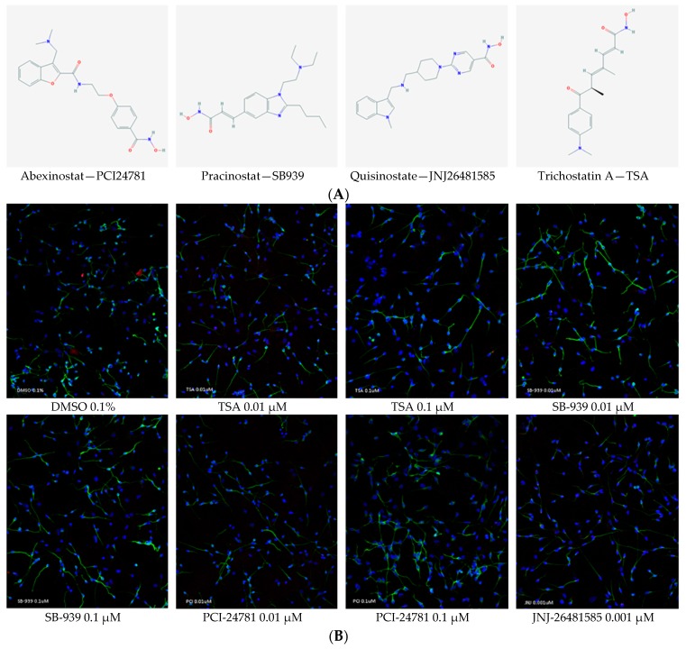
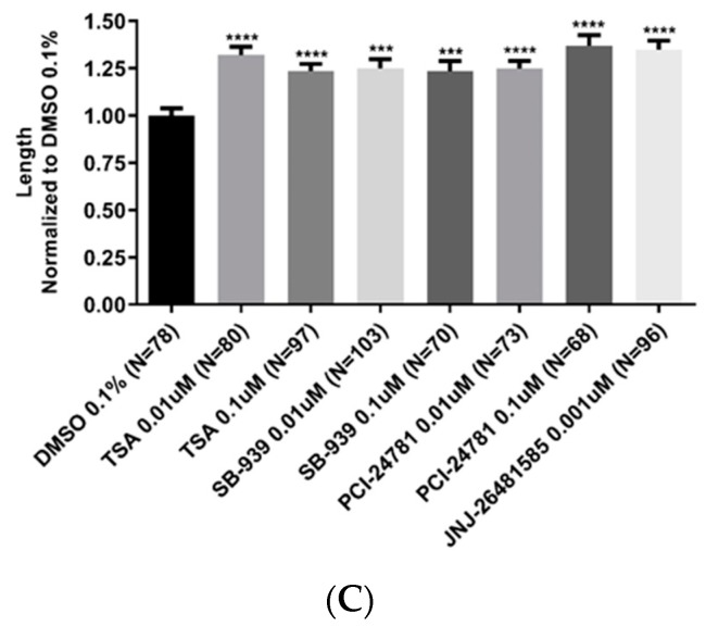
Figure 5.
(A) Chemical structures of the test compounds; abexinostat (PCI24781), pracinostat (SB939), quisinostat (JNJ26481585), and trichostatin A (TCA). (B) Representative fluorescent images (20× magnification) for each treatment. Human neural progenitor cells were differentiated for 5 days; then treated for 24 h with 0.1% DMSO (as control), and trichostatin A, JNJ26481585, SB939, and PCI24781 (as test compounds). Then immunostaining was performed with neuronal-specific β-tubulin III antibody (green) to visualize neuronal processes and quantify neurite lengths and Dapi (blue) to visualize cell bodies. (C) Statistical analysis of neurite length in various groups. As depicted, all test compounds were able to induce neurite outgrowth: PCI24781 (0.1 µM) had the greatest effect with 37% increase compared with that of the 0.1% DMSO. It was followed by JNJ26481585 (0.001 µM) with 34% increase. Trichostatin A was used as a positive control. Quantitative analysis of neurite length was done using ImageJ software. Two fields of view were analyzed per well. Two biological replicates were used per treatment and two independent experiments were done. Error bars represent SEM; *** p < 0.001, **** p < 0.0001.


