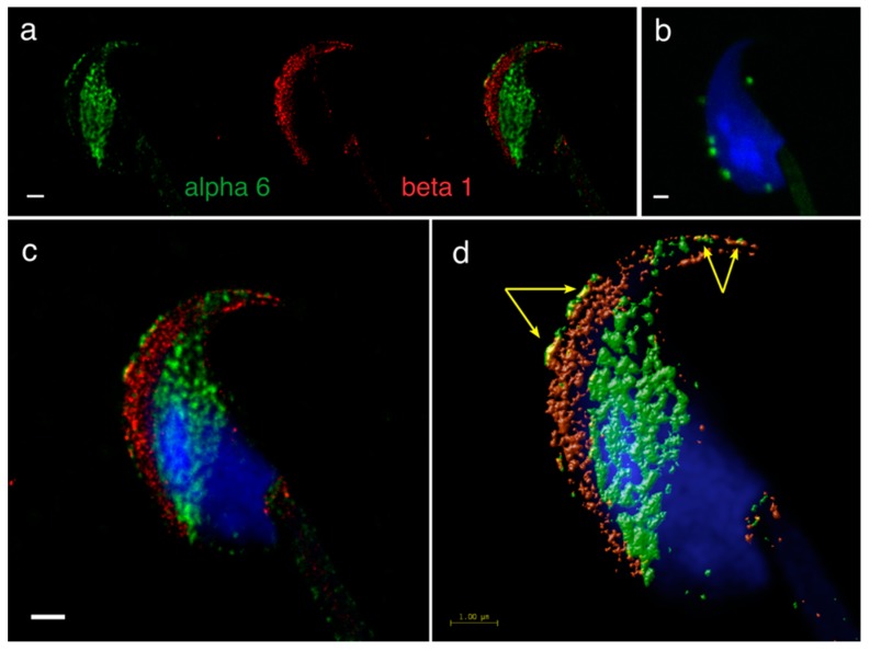
Figure 5.
Mutual localization of α6 and β1 subunit and the presence of α6β1 heterodimer reveals by STED (Stimulated Emission Depletion) super-resolution microscopy and PLA. (a) α6 (green) is localized in plasma membrane over the acrosomal cap, apical hook and equatorial segment in contrast to β1 (red) localized in plasma membrane overlying the acrosomal cap stretching to apical hook and in outer acrosomal membrane. (b) PLA confirmed the presence of the α6β1 heterodimer in mutual places such as the plasma membrane over the acrosomal cap and apical hook. (c) STED dual-color imaging present overlay of both subunits. (d) Huygens software was used for better visualization of colocalization area (yellow, pointed by yellow arrows). Colocalization maps were based on Pearson’s correlation coefficient. Nucleus is visualized with Dapi (blue). Scale bar represents 1 μm (a–d).

