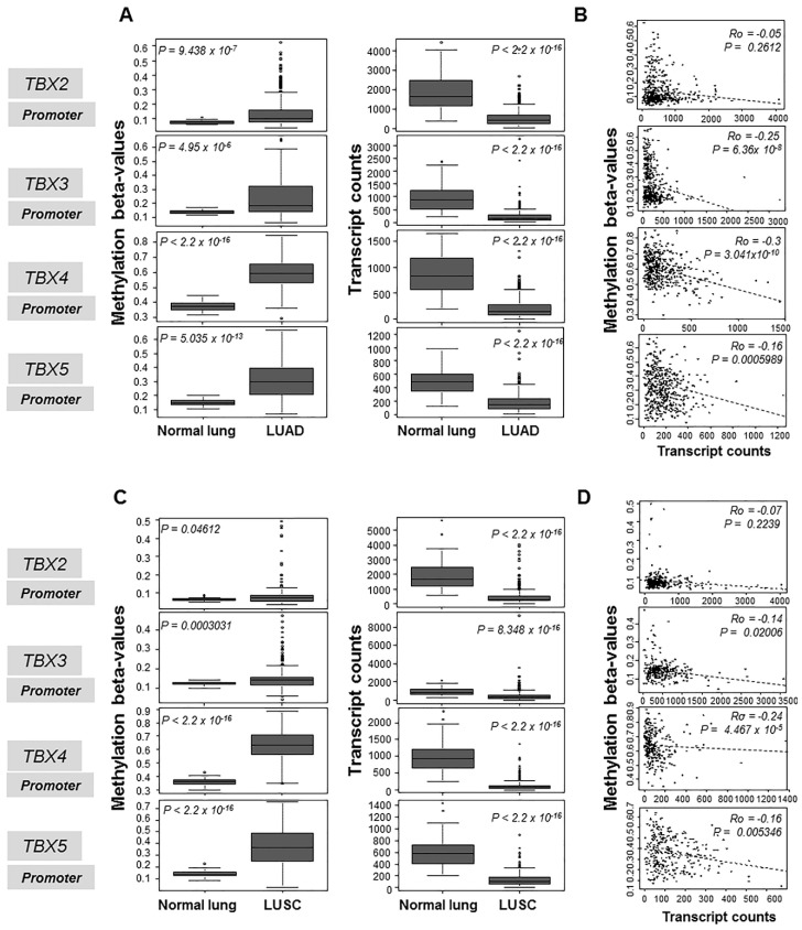
Figure 1.
Increased promoter-level methylation of the TBX2 subfamily in human LUADs and LUSCs compared to normal lung tissues. Promoter-level methylation β-values (left panels) and mRNA levels (right panels) in 460 LUADs (A,B) and 370 LUSCs (C,D) were propagated from the MethHC database of methylation and gene expression in cancer. Methylation β-values and mRNA levels of the TBX genes were statistically analyzed by the Wilcoxon rank sum test in LUADs relative to normal lung tissues (A). Methylation β-values and mRNA levels of the TBX genes were also similarly statistically analyzed LUSCs relative to normal lung tissues (C). Methylation β-values and mRNA expression levels for each of the four TBX genes were statistically correlated in LUADs (B) and in LUSCs (D) using Spearman’s correlation. Statistical analyses and plots were performed and generated using the R language and environment.

