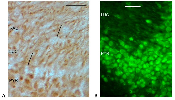
Figure 2.
Immunohistochemical localization of GluK1 (A) and GluK2 (B) antibodies in the CA3 region of the murine hippocampus. The immunohistochemical picture suggests mainly postsynaptic GluK1 and GluK2 localization. PYR: pyramidal layer; LUC: stratum lucidum; RAD: stratum radiatum. Arrows on Figure 2A point to unstained mossy fiber terminals. See Appendix A for methods. Bars: 50 µm.

