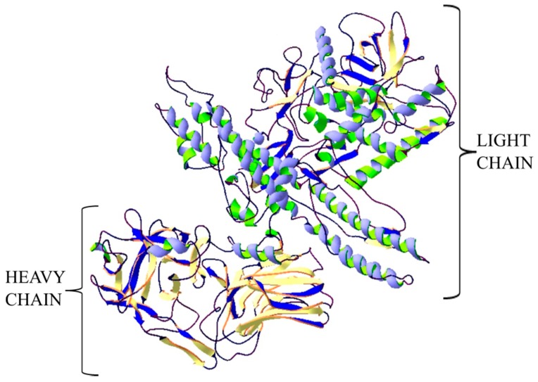
Figure 1.
Structure of botulinum neurotoxin type A. The crystal structure of protein (PDB: 3BTA) [28] was taken from the RCSB PDB databank (http://www.rcsb.org/) (access on 05.03.2018). Visualization of the three-dimensional structure of the protein was performed using the Swiss-PdbViewer (http://spdbv.vital-it.ch/) (access on 05.03.2018.) [29].

