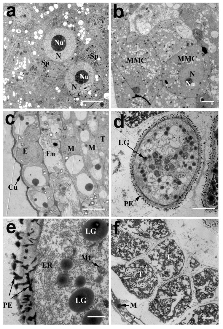
Figure 3.
Transmission electron micrographs of cross-sections through anthers of N. nucifera ‘Honghu Hong’. (a) The sporogemous cells at the sporogenous cell stage. (b) The microspore mother cell at the MMC stage. (c) The anther wall comprising the epidermis, endothecium and middle layers at the MMC stage. The arrow indicates the epidermal cuticle. (d) The young microspores at the early young microspore stage, the arrows indicate the lipid granule and primexine respectively. (e) The magnification image of the young microspore wall at the early young microspore stage, the arrows indicate the primexine and mitochondria, respectively. (f) The magnification image of the tapetal cells at the early young microspore stage, the arrow indicates the middle layer. Cu, anther epidermal cuticle; E, epidermis; En, endothecium; ER, endoplasmic reticulum; LG, lipid granule; M, middle layer; MMC, microspore mother cell; Mt, mitochondria; N, nucleus; Nu, nucleolus; PE, primexine; SP, sporogenous cell; T, tapetum. (a–d,f) Bar = 5 μm; (e) Bar = 1 μm.

