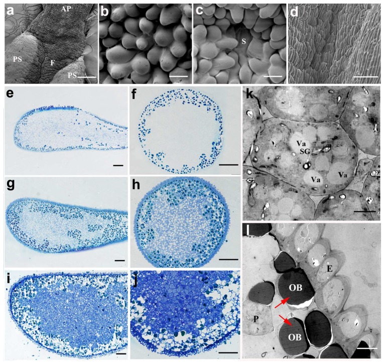
Figure 6.
The structure and development of the anther appendage of N. nucifera ‘Honghu Hong’. (a) View of the appendage–anther junction. (b) The surface structure of the appendage at the mature pollen stage. (c) The surface structure of the filament at the mature pollen stage. (d) The surface structure of the anther at the mature pollen stage. (e–j) Longitudinal and lateral sections of the intact appendage at the MMC stage (e,f), the young microspore stage (g,h) and the mature stage (i,j). (k) The transmission electron micrograph of a parenchyma cell, the white arrows indicate the starch granules. (l) The transmission electron micrograph of the epidermis, the red arrows indicate the osmiophilic body in the parenchyma cells. AP, appendage; E, epidermis; F, filament; OB, osmiophilic body; P, parenchyma; PS, pollen sac; S, stomate; SG, starch granule; Va, vacuole. (a) Bar = 300 μm; (b,c) Bar = 10 μm; (d) Bar = 50 μm; (e–j) Bar = 100 μm; (k) Bar = 5 μm; (l) Bar = 10 μm.

