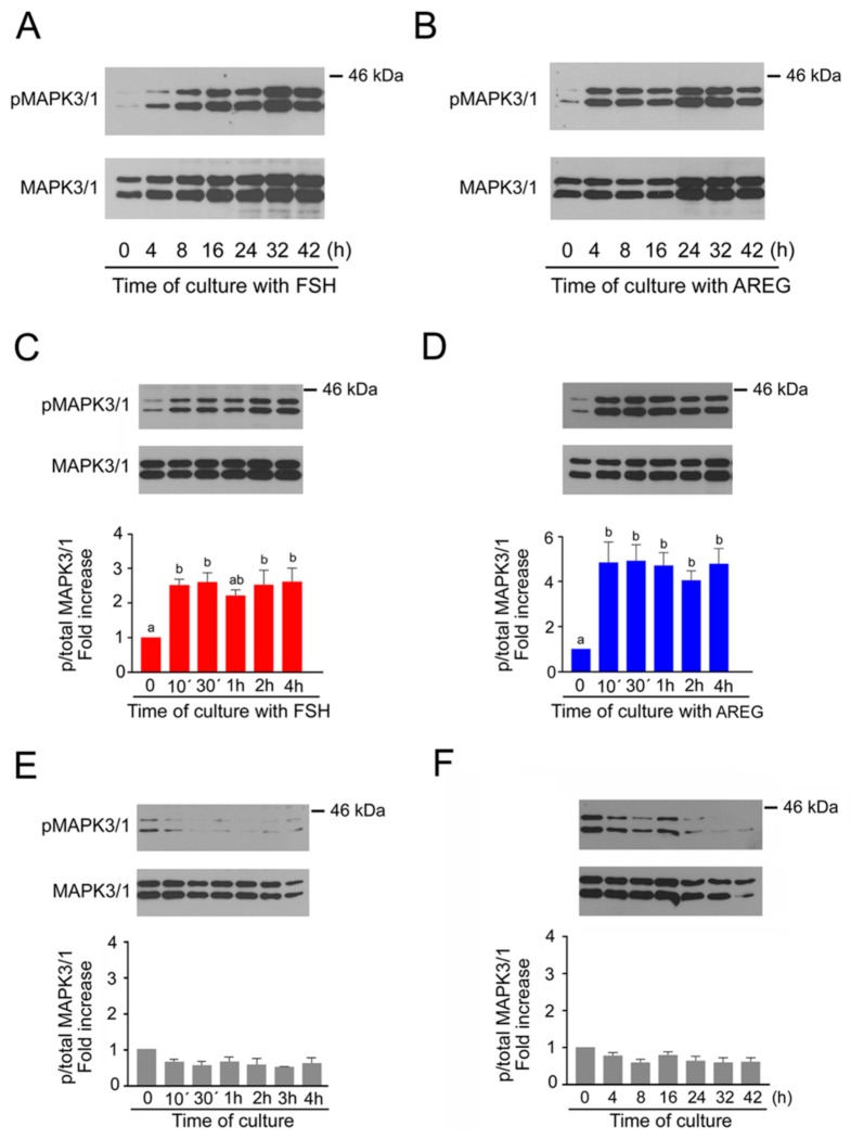
Figure 1.
Time course of MAPK3/1 activation in cumulus-enclosed oocytes (COCs) stimulated with follicle-stimulating hormone (FSH) or amphiregulin (AREG). The panels show representative results of immunoblotting of phosphorylated and total MAPK3/1 in samples of 25 COCs cultured in vitro for the indicated periods of time. The experiments shown in panels A and B were repeated twice with the same results. The experiments shown in panels (C–F) were repeated three times. Quantification of the activated MAPK3/1 was performed by densitometry and is shown in the graphs as proportions of phosphorylated and total MAPK3/1 and expressed in arbitrary units as the fold increase over the proportion found in unstimulated COCs at the beginning of the culture. (A) Activation of MAPK3/1 in FSH-stimulated COCs during the long-term culture. (B) Activation of MAPK3/1 in AREG-stimulated COCs during the long-term culture. (C) Activation of MAPK3/1 in FSH-stimulated COCs during the short-term culture. (D) Activation of MAPK3/1 in AREG-stimulated COCs during the short-term culture. (E) Activity of MAPK3/1 in COCs cultured for short-term period in control medium without FSH and AREG. (F) Activity of MAPK3/1 in COCs cultured for long-term period in control medium without FSH and AREG. The different superscripts above the columns indicate significant differences (p < 0.05).

