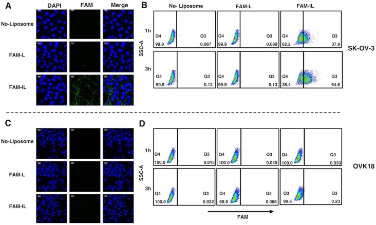
Figure 4.
Immunoliposome enhanced cellular uptake in CD44 postive cells. (A,C) Confocal Microscopy image after 2 h treatment FAM-L and FAM-IL, Each scale bar shows 20 µm. (B,D) Flow cytometry analysis after 1 h and 3 h treatment FAM-L and FAM-IL. FAM-L and FAM-IL were evaluated for the cellular uptake in SK-OV-3 (A,B) and OVK18 (C,D), SSC-A is side scatter area. Data are representative of three replicates.

