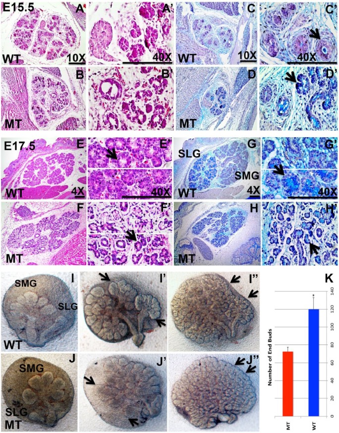
Figure 2.
Hematoxylin and eosin, alcian blue, and periodic acid–Schiff staining of submandibular glands in wild-type (WT; A, C, E, G series) and Irf6-null (mutant type [MT]) embryos (B, D, F, H series) and ex vivo culture of salivary gland explants. At embryonic day 15.5 (E15.5), histologic staining shows round ductal cells with a single circular lumen per duct (A, C series; black arrow). In comparison, the staining in Irf6-null glands shows significant ductal cell disorganization and disruption of ductal lumens (arrow in D′). At E17.5, the serous and mucous acini are detected in WT gland (black arrow in E′; E, G series), while the mucous acini (black arrow) cannot be distinguished in Irf6-null salivary glands and the interstitial spaces among the acini are wider when compared with WT (F, H series; black arrow in H′). In salivary gland explants, the sublingual gland (SLG) and submandibular gland (SMG) are formed in the WT and Irf6-null littermates at E13.5 (I, J). Explants show branched SLG and SMG after 2 d (I′, black arrow) and a remarkable number of end buds (black arrow) and branching by day 4 (I′′) of cultivation. In Irf6-null explants, the end buds of SLG and SMG seem fused by day 2 (J′, arrow) of incubation, and by day 4, many end buds appear to be coalesced or not separated (J′′, arrow). The number of end buds was counted 4 d post-incubation in 3 biological replicates, and it is significantly decreased in Irf6-null embryos (MT) versus WT littermates (WT; K). The values are the mean number of end buds, and the bars represent the standard deviation. Asterisk represents statistical significance of WT versus Irf6-null samples, P < 0.05. Scale bars are 150 μm.

