Fig. 5.

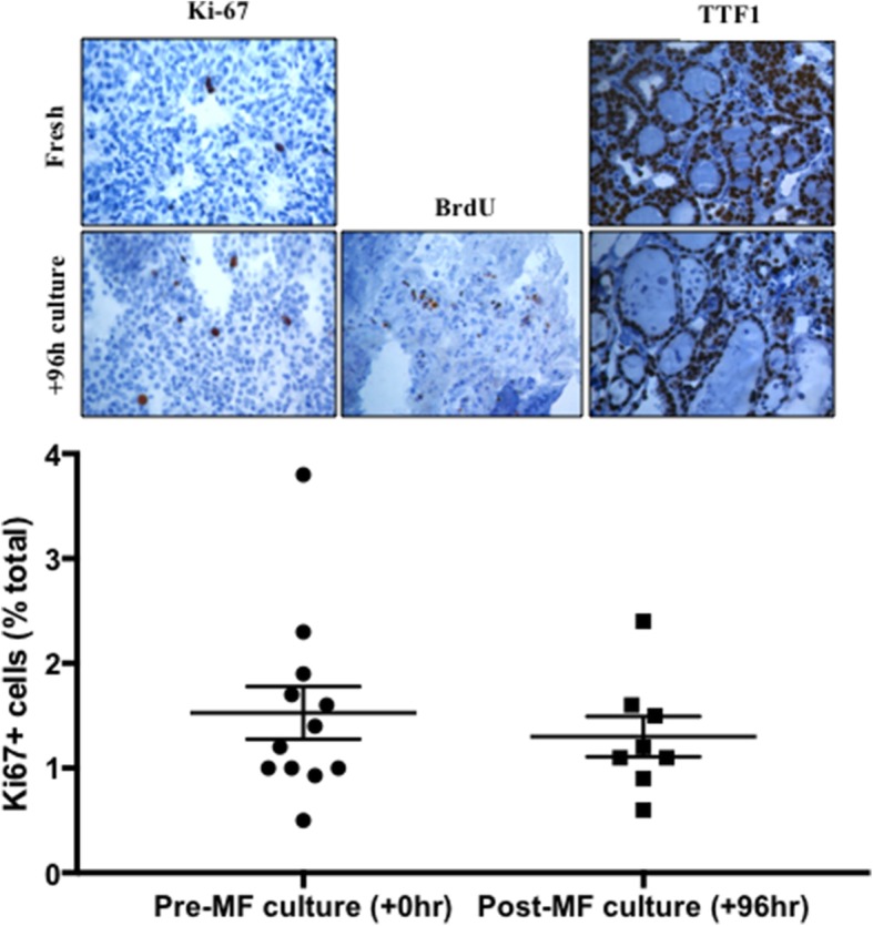
Percentage Ki-67 positivity in fresh (+ 0 h) and post-culture (+ 96 h) thyroid tumour tissue. Mean Ki-67 positivity was 1.52 ± 0.52% and 1.3 ± 0.34% in fresh and post-culture tissue, respectively; no significant difference was observed (n = 8; p = 0.32 [paired t-test]). Inset: Representative photomicrographs of Ki67, BrdU and TTF1 positive brown nuclei and counterstained with haematoxylin (× 400). BrdU staining is only shown in cultured tissue due to the requirement of perfusion with the molecule in order to elicit its incorporation and subsequent detection; 12.09 ± 4.38% cells stained positive for BrdU incorporation (n = 3)
