Fig. 1.
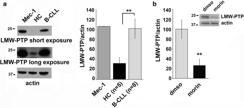
LMW-PTP phosphatase expression is downregulated by morin treatment in Mec-1 cells. a Left: Immunoblot analysis with anti-LMW-PTP antibodies from lysates of Mec-1 cells and primary B cells purified from peripheral blood of a representative healthy control (HC) or CLL patient (B-CLL). The stripped filters were reprobed with anti-actin antibodies as loading control. Right: quantification by laser densitometry of the protein bands. Each sample was normalized to the relative actin and data are expressed as percentage (value of Mec-1 cells set as 100). The quantification of protein levels in Mec-1 cells are relative to three independent experiments. For primary B cells the number of samples is HC n = 5 and B-CLL n = 9. Data are expressed as mean ± SD. b Quantification by laser densitometry of the LMW-PTP protein levels normalized to the respective actin in Mec-1 cells treated with 50 μM morin or DMSO as control for 24 h. A representative immunoblot analysis is shown on the top of the panel. The quantifications are relative to three independent experiments (n = 3). Error bars, SD. **p ≤ 0.01

