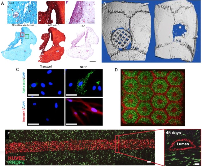
Figure 3.

Bioprinted tissues. (A): 3D‐bioprinted chondrocyte‐derived iPSCs at week 5 of differentiation, sections stained for GAGs, Safranin O for cartilage (with nuclear counterstain), and H&E for extracellular matrix (with nuclear counterstain) (the scale bar represents 100 μm or 500 μm) (Reproduced with permission from 58). (B): Micro‐CT images of polylactic acid/hydroxyapatite scaffold (left) versus bone defect without scaffold (right) after 4 weeks in vivo. (Adapted and reproduced with permission from 58). (C): Immunocytochemistry of MSCs for cardiac proteins in the transwell versus nanothin and highly porous membrane methods (Adapted and reproduced with permission from 59). (D): 3D bioprinting of hydrogel based hepatic construct. Images (×5) showing patterns of fluorescently labeled hiPSC‐HPCs (green) in 5% (wt/vol) GelMA and supporting cells (red) in 2.5% (wt/vol) GelMA with 1% GMHA on day 0 (Scale bars, 500 µm; Reproduced with permission from 60). (E): Top‐down (left) and cross‐sectional (right) confocal microscopy images of bioprinted vascular networks supporting human neonatal dermal fibroblast‐laden (green) matrix and HUVEC (red) lined channels obtained after 45 days of perfusion culture. (Scale bar: 100 µm; Reproduced with permission from 61). Abbreviations: DAPI, 4′6‐diamidino‐2‐phenylindole; H&E, hematoxylin and eosin; HNDFS, human neonatal dermal fibroblasts; HUVECs, human umbilical vein endothelial cells; NTHP, nanothin and highly porous.
