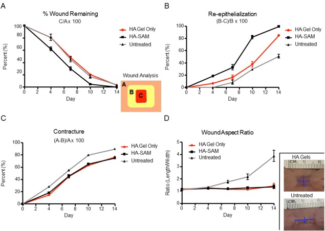
Figure 4.

Quantitative Analysis of Wound Healing. (A): Percentage (%) wound remaining was calculated by dividing the area of the remaining wound by the original wound size (C/A × 100). HA‐SAM groups had significant acceleration of wound closure times resulting in wound closure 3–4 days before other groups. (B): Wound re‐epithelialization was calculated by measuring newly re‐epithelized skin, taking into consideration remaining wound area and contraction ([B‐C]/B × 100). HA‐SAM groups had significantly greater wound re‐epithelialization at all time‐points compared with other groups. (C): Contraction was measured based on original wound size and the area of re‐epithelized skin ([A‐B]/A × 100). HA only and HA‐SAM groups had significantly less contraction compared with untreated animals. (D): Wound aspect ratio was determined to describe observed changes in the shape and direction of wound contraction between groups (length:width). HA only and HA‐SAM groups displayed symmetrical contraction, with aspect ratios close to 1, while other groups showed asymmetrical contraction with aspect ratios closer to 4.
