Figure 6.

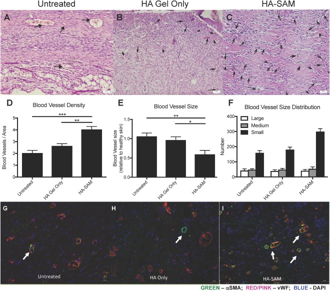
Blood Vessel Formation in Healing Wounds. H&E stained sections of wound area demonstrating blood vessels within the newly regenerated untreated (arrows) (A), HA only (B), or HA‐SAM‐treated (C) skin. (D): Blood vessel density within areas of regenerated skin. There were significantly more blood vessels in HA‐SAM‐treated animal compared to HA only and untreated groups. Counts were performed on six representative fields of view. (E): Blood vessel area was calculated and represented as relative to blood vessel size within healthy skin of the same mouse. Average blood vessel size was significantly smaller in HA‐SAM‐treated animal compared to HA only and untreated groups. (F): HA‐SAM‐treated wounds had similar density of large or medium sized blood vessels, but significantly greater density of small blood vessels compared to HA only‐treated and untreated wounds. Immuno‐fluorescent staining for αSMA and vWF to stain for blood vessels in the regenerating skin of untreated (G), HA only (H), and HA‐SAM (I) animals. HA‐SAM samples had significantly more blood vessels than other groups. These blood vessels also appeared smaller in size than vessels within skin from other groups. Scale bars = 50 µm (A–C) Scale bars = 25 µm (G–I). (*, p < .05; **, p < .01; ***, p < .01). Abbreviations: αSMA, α‐smooth muscle actin; DAPI, 4′,6‐diamidino‐2‐phenylindole; HA, hyaluronic acid; SAM, solubilized amnion membrane; vWF, von Willebrand factor.
