Figure 7.

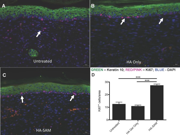
Cell Proliferation in Healing Wounds. Immuno‐fluorescent staining for proliferating cells in the epidermis of untreated (A), HA only (B), and HA‐SAM (C) animals. Antibodies for keratin 10 were used to highlight the epidermis and Ki‐67 to stain proliferating cells. More proliferating cells were identified in the basal layer of the epidermis of HA‐SAM‐treated animals compared to HA‐gel only‐treated and untreated groups (arrows). In untreated groups proliferating cells were also identified within the dermis, while this was rare in HA only and HA‐SAM groups. Significantly more proliferating cells were observed in HA‐SAM‐treated animals compared to HA‐only‐treated and untreated groups. Scale bars = 25 µm. (***, p < .0001). Abbreviations: DAPI, 4′,6‐diamidino‐2‐phenylindole; HA, hyaluronic acid; SAM, solubilized amnion membrane.
