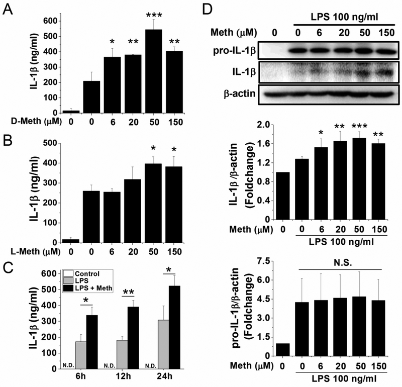
Fig. 2.
Meth treatment potentiated LPS-induced IL-1β processing and release. Microglia were pretreated 6 h with LPS (100 ng/ml) and then incubated 12 h with different concentrations of D-Meth (A) or L-Meth (B). Alternatively, after 6 h pretreatment of LPS, cells were incubated with Meth (50 μM) for indicated times (C). The supernatants were collected, and concentrations of secreted IL-1β were measured by ELISA. The optimized time point (12 h) and concentration (50 μM) of Meth treatment were applied on cultured microglia. The production and processing of IL-1β were detected by western blot (D). After standardized with β-actin, the densitometry of bands was analyzed and quantified, as shown in bar graphs (D). * p < .05, ** p < .01, *** p < .001 vs LPS;

