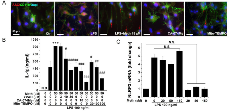
Fig. 4.
Meth-induced inflammasome activation was inhibited by lysosomal and mitochondrial ROS-specific inhibitors. Punctate ASC immunoreactivity was detected in the cytoplasm of Meth (18 μM) treated microglia (A). Microglia pretreated with lysosomal cathepsin B inhibitor (CA-074Me 10 μM) and mitochondrial ROS inhibitor (Mito-TEMPO 100 μM) exhibited minimal ASC immunoreactivity (A). Blue: DAPI; Green: CD11b; Red: ASC. Isolated microglia were pretreated with different concentrations of inhibitors before LPS and Meth application; the secreted IL-1β were quantified by ELISA (B). The transcriptional level of NLRP3 was measured by qPCR on Meth-treated microglia with or without priming signal (C). *** p < .001 vs LPS; # p < .05, ## p < .01, ### p < .001 vs LPS + Meth 50 μM. Scale bar, 50 μm.

