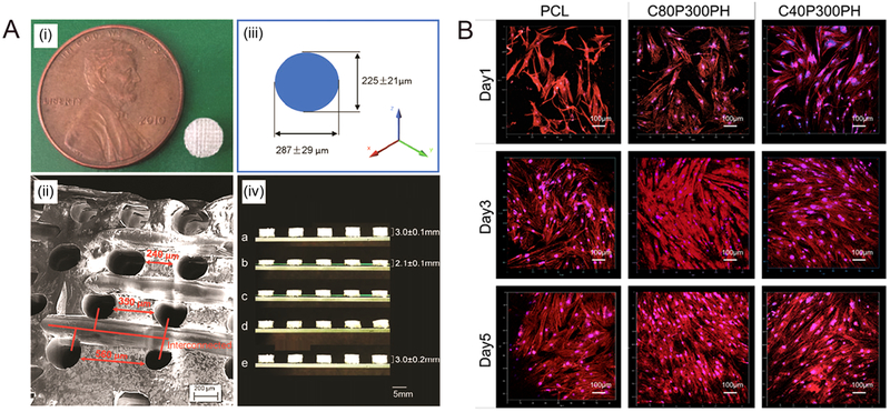
Figure 4.

(A) The fabricated scaffold. (i) A 5-mm-diameter and 3-mmthickness scaffold compared to a cent. (ii) The SEM image of the pore distribution in the scaffold. (iii) Varied pore diameters in different directions. (iv) The potential for minimally invasive application; a, sample original shape; b, temporary shape at −18 °C; c,0 s at 37°C; d, 10 s at 37 °C; e, 3 min at 37°C. (B) Confocal microscopy images of MSC growth and spreading morphology on printed samples of C40P300PH and C20P300PH when compared with PCL control after 1-, 3-, and 5-day culture. The color red represents cell cytoskeleton and the color blue represents cell nuclei.
