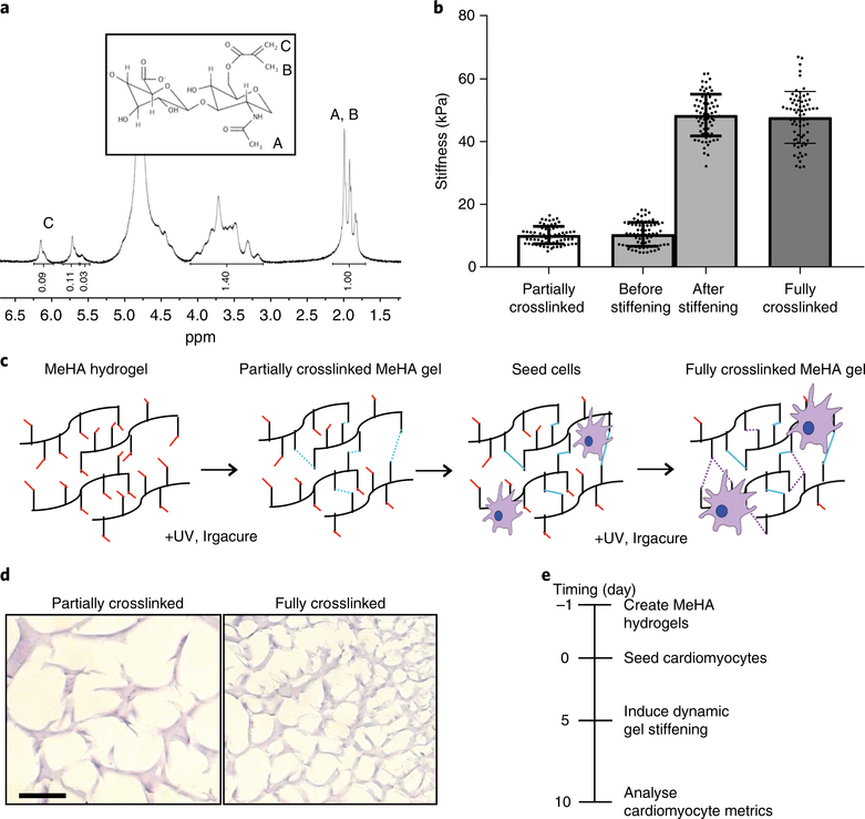
Fig. 1 |. MeHA synthesis and schematic of dynamic stiffening.
a, NMR spectrum of 50 kDa hyaluronic acid after methacrylate functionalization (the degree of methacrylation was around 40%). Inset, the MeHA structure. b, Plot of atomic-force-microscopy-measured stiffness for hydrogels of ‘partially crosslinked’ (10 kPa or ‘physiological’), ‘stiffened’ (a hydrogel originally crosslinked to 10 kPa before additionally stiffened to 50 kPa) and ‘stiff’ (50 kPa) MeHA (n = 70 measurements). Data are mean ± s.d. with individual points. c, Schematic illustrating cell seeding on soft MeHA substrates followed by sequential, in situ dynamic stiffening. d, MeHA hydrogels stained with haemotoxylin and eosin to visualize crosslinking. Scale bar, 10 μm. e, Timeline indicating cell seeding, dynamic stiffening induction and analysis.

