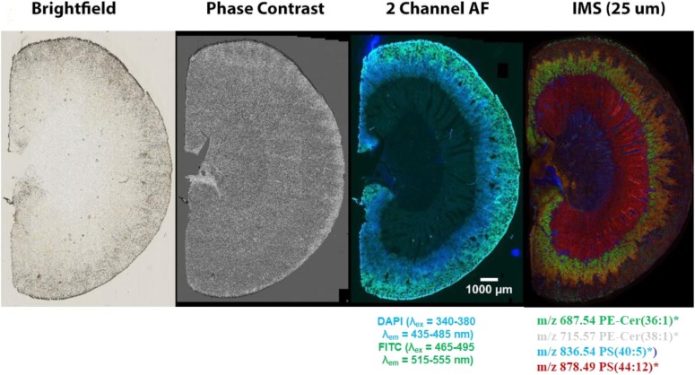
FIGURE 3.
Images of a mouse kidney section taken with different imaging modalities. The combination of the fluorescence image, which clearly shows tissue morphology, with IMS images, which gives unparalleled molecular specificity, is an excellent combination for image overlay to extract maximum spatial molecular information from discrete areas within the tissue.

