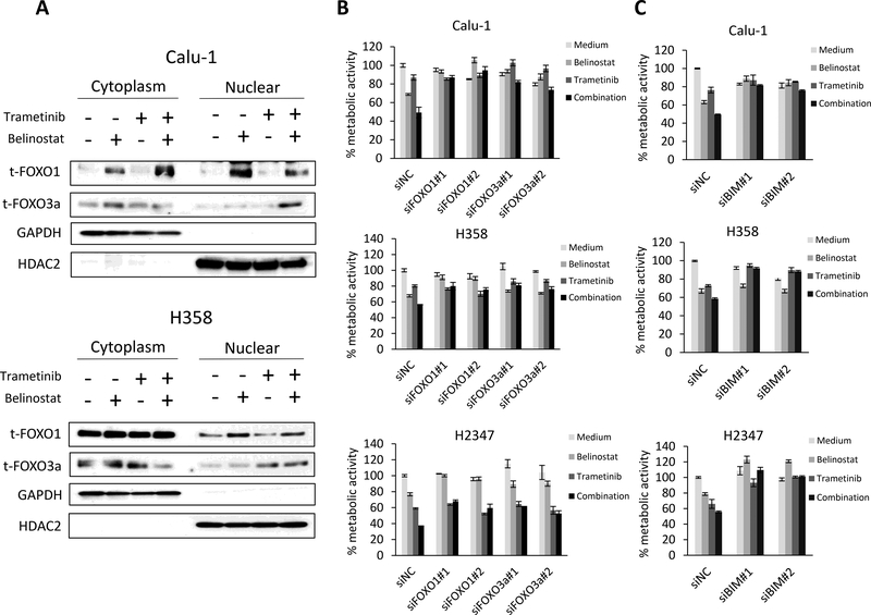
Figure 3. FOXO1 and FOXO3a protein levels increase in the nuclear fraction with combination treatment and are responsible for the increased cell death through regulation of BIM.
(A) Tumor cells were treated with belinostat (1000 nmol/L) and/or trametinib (100 nmol/L) for 4 h. The cells were lysed to extract nuclear and cytoplasmic fractions using the NE-PER Nuclear and Cytoplasmic Extraction Reagents and the indicated proteins were detected by immunoblotting. The results shown are representative of 3 independent experiments. (B) Control or FOXO1- or FOXO3a-specific siRNAs were introduced into Calu-1, H358, and H2347 cells. After 24 h, the cells were incubated with belinostat (100 nmol/L) and/or trametinib (10 nmol/L) for 72 h and metabolic activity was determined by MTT assays. FOXO1 or FOXO3a knockdown was confirmed by immunoblotting (Supplementary Fig. 4). The percentage of metabolic activity is shown relative to untreated controls. Each sample was assayed in triplicate, with each experiment repeated at least 3 times independently. (C) Control or BIM-specific siRNAs were introduced into Calu-1, H358, and H2347 cells. After 24 h, the cells were incubated with belinostat (100 nmol/L) and/or trametinib (10 nmol/L) for 72 h and lung cancer cell metabolic activity was determined by MTT assays. BIM knockdown was confirmed by immunoblotting (Supplementary Fig. 4). The percentage of metabolic activity is shown relative to untreated controls. Each sample was assayed in triplicate, with each experiment repeated at least 3 times independently.

