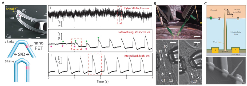
Figure 5. 3D Si nanowire FETs for cardiac recording.
(A) Kinked nanowire probe for intracellular recording, showing an SEM image of a device (upper left), schematic diagrams of the material design (lower left), and representative electrical recordings (right). During the experiment, extracellular recording (I) was obtained, followed by the transitions (II) into internal recording until it maintained steady-state intracellular recording (III). Scale bar, 5 µm. (B) Multiplexed recording is possible by using two independent nanoFETs mounted over micromanipulators (upper). Two probes, P1 and P2, were positioned at two adjacent cardiomyocytes (lower left). Alternatively, they were placed in two different subcellular locations of the same cardiomyocyte (lower right). Scale bars, 1 cm (upper) and 10 µm (lower). (C) A schematic illustration (upper) and an SEM image (lower) of a branched nanotube-enabled nanoFET intracellular recording device, where the intracellular fluid can be introduced to the nanoFET by a silicon dioxide nanotube. (A) Reproduced with permission.[11] Copyright 2010, AAAS. (B) Reproduced with permission.[12] Copyright 2014, Springer Nature. (C) Reproduced with permission.[13] Copyright 2012, Springer Nature.

