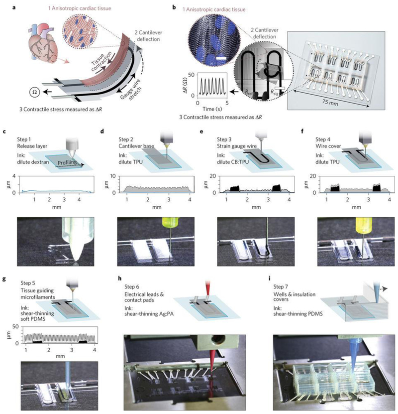
Figure 2. Printed cell-integrated electronics.
a, Device working principle. When an anisotropic engineered cardiac tissue contracts, it will deflect a cantilever substrate and stretch a soft strain sensor embedded in the cantilever. The contractile stress of the tissue can then yield resistance change in the sensor. b, Images showing a fully printed final device. Inset 1: Confocal microscopy image of immunostained cardiac tissue on the cantilever surface. Blue, DAPI nuclei stain. White, α-actinin. Inset 2: An image of a cantilever deflecting upon tissue contraction. Inset 3: Representative resistance signal from the embedded strain sensor. c–i, Schematic diagrams and photos showing the automated printing of the device on a glass slide substrate in seven sequential steps. A stylus profiling cross-sectional contour of the cantilever is also shown for steps 1–5. For details of the printing, please refer to ref 30. Figures are reproduced with permission from ref 30. Copyright 2017 Nature Publishing Group.

