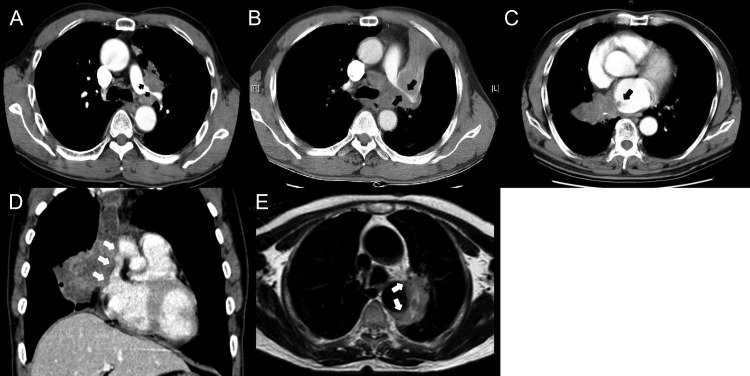
Fig. 2.
Representative images of great vessel invasions (GVIs): (A) irregular indentation at the tumor and left pulmonary artery contact border (black arrows), (B) slit-like narrowing of left pulmonary artery contact border by the tumor (black arrows), (C) presence of intra-luminal mass formation (black arrows), (D) tumor contacting >5 cm of adjacent great vessel and obliteration of the intervening fat plane between tumor and adjacent great vessel (white arrows), and (E) tumor contacting more than half of the circumference of the aortic wall (white arrows).

