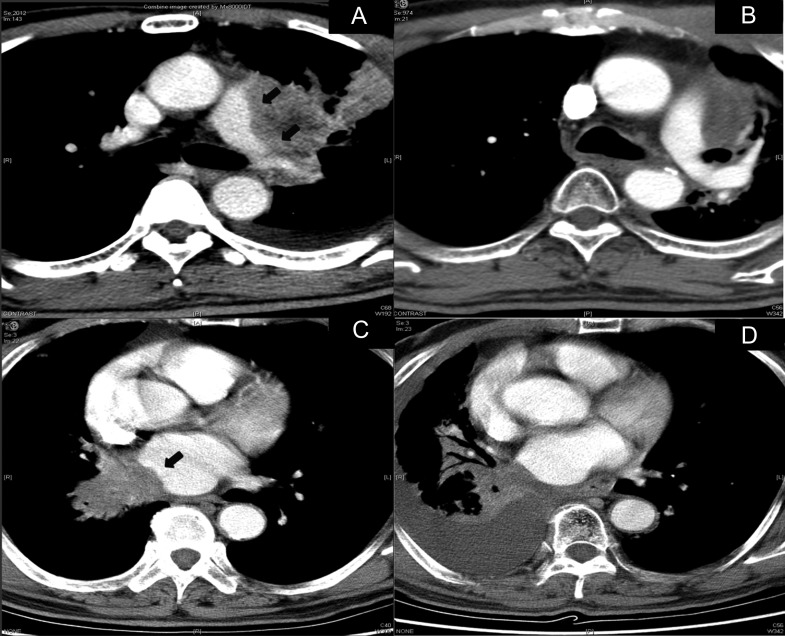
Fig. 5.
Computed tomography (CT) scans of patients who experienced critical events. (A) CT scan of 68-year-old male patient with tumor invading left pulmonary artery (black arrows) before concurrent chemo-radiotherapy (CCRT), (B) CT scan at 17 months after CCRT, when he experienced hemoptysis. There was no evidence of any left pulmonary artery rupture. (C) CT scan of 66-year-old male patient with tumor invading heart (black arrows) before CCRT, (D) CT scan at 2 months after the onset of hemoptysis at 2 months after CCRT. There is no evidence of any rupture in the heart.

