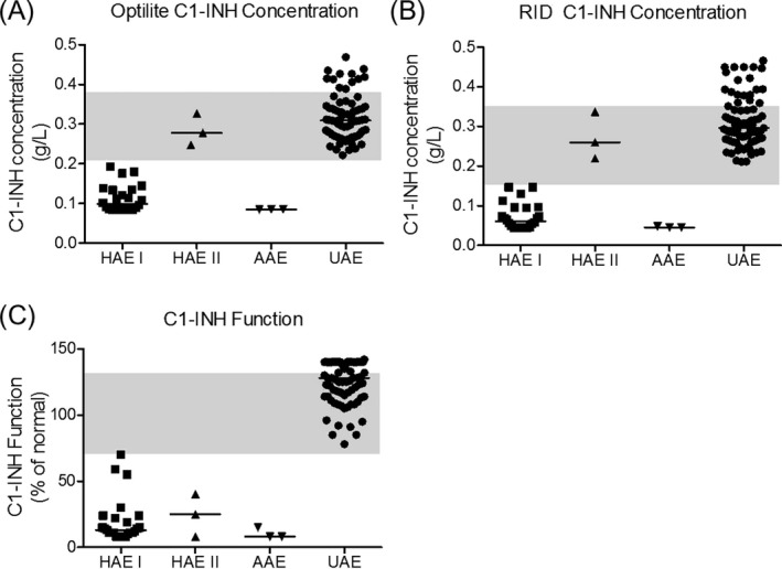
Figure 5.

C1‐INH concentration and C1‐INH functional activity in clinical samples. The C1‐INH concentrations were measured using the Optilite assay (A) and radial immunodiffusion (RID, B). The functional activity of C1‐INH is also shown (C). Shaded areas represent the normal range of the assays
