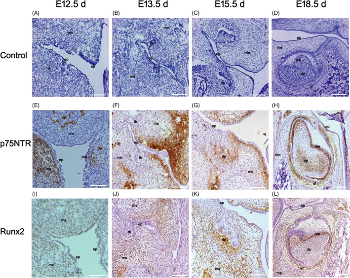
Figure 1.

The results of HE staining (A‐D) and immunohistochemistry staining (E‐L). The rat original oral epithelium(ep) invaginated to form dental lamina at E12.5 d (A). The dental germs turned up and entered the bud stage at E13.5 d, the cap stage at E15.5 d and the bell stage at E18.5 d (B‐D). p75NTR immunoreactivity was little expressed at E12.5 d in some subjacent mesenchyme area under the oral pit epithelium (E). p75NTR immunoreactivity was enhanced at E13.5 d, but not detected around the tooth buds(tb) (F). At the cap stage, p75NTR began to express in the mesenchyme(me) of dental papilla(dp), liking that p75NTR‐positive ectomesenchymal cells migrate to dental papilla from the oral pit (G). At the bell stage (H), the p75NTR expression became weak in dental papilla and strong in dental follicle(df), but firstly expressed in the inner enamel epithelium(iee). Runx2 was hardly detected at E12.5 d (I) and became weakly positive in the mesenchyme around the tooth buds at E13.5 d (J). It was positive in mesenchyme area and became significantly enhanced under epithelial–mesenchymal interaction of the tooth germs at E15.5 d, but not positive in the mesenchyme area under the oral pit epithelium (K). At the bell stage of E18.5 d, Runx2 was positive in the inner enamel epithelium and dental follicle area but little in the dental papilla (L). Scale bar represents 100 μm
