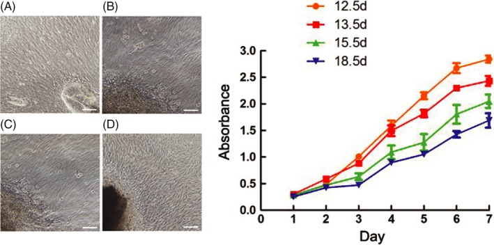
Figure 2.

The cell culture and proliferation assay in vitro. All the cells exhibited a fibroblast‐like morphology, and no difference was observed between the cells from different tooth development stages (A‐D). The CCK‐8 assay showed that the cell proliferation capacity decreased during the development proceeding (E). But there was also no significant difference (P > 0.05). Scale bar represents 100 μm
