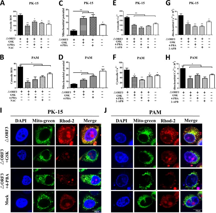
FIG 6.
Blocking UPR or ER stress by chemical inhibitors decreased cellular ROS levels, increased MMP, and reduced cytosolic and mitochondrial Ca2+ levels in PK-15 and PAM cells infected with ORF3-deficient PCV2. PK-15 and PAM cells were incubated in 24-well plates. Cells were infected with ΔORF3 (MOI of 1), treated with 5 μM GSK or 2 mM 4-PBA at 2 hpi, and then incubated for an additional 46 h. The chemicals 2-APB (50 μM, as a Ca2+ channel IP3R inhibitor) and NAC (10 mM, as a ROS scavenger) were used as controls. (A and B) The changes in cytosolic ROS levels in PK-15 and PAM cells were evaluated according to the mean values of DCF fluorescence intensity. (C and D) JC-1 fluorescence is shown as the ratio between red and green fluorescence signals. (E to H) The changes in cytosolic and mitochondrial Ca2+ levels were evaluated according to the mean values of fluo-3 and rhod-2 fluorescence intensity. (I and J) By confocal microscopy, serial treatments were followed with 5 μM rhod-2 for 30 min, 100 nM MitoBright Green dye for 15 min, and DAPI for 10 min in the dark after 48 hpi. Blue, nucleus; green, mitochondrial tracker; red, mitochondrial calcium tracker. Scale bars, 10 μm. Data in the bar charts in panels A to H represent the mean ± SEM of three independent experiments. *, P < 0.05; **, P < 0.01.

