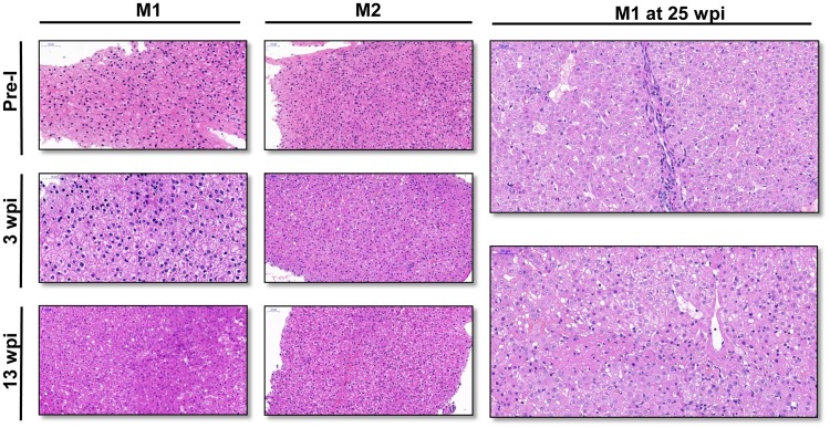
FIG 5.
Histopathology of liver tissue in monkeys. (A) Liver biopsy specimens were collected from M1 and M2 1 week before inoculation (Pre-I), at 3 weeks postinoculation (wpi), and at 13 wpi. Hydropic degeneration and slight inflammatory cell infiltrates were found in biopsy specimens at 3 wpi for both monkeys. Focal necrosis of hepatic cells and dilation of the liver sinusoid were seen in the liver biopsy specimen of M1 at 13 wpi. (B) Liver tissues were collected at 25 wpi from M1, showing focal necrosis around the central vein and lobular inflammatory cell infiltrates. Ground-glass hepatocytes were also scattered in the liver. Bars, 50 μm.

