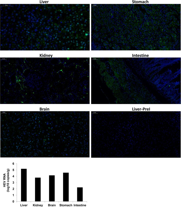
FIG 6.
Extrahepatic replication of HEV in monkey M1. The viral RNA and ORF2 protein of HEV was detected using quantitative real-time PCR and an immunofluorescence assay, respectively, in liver, kidney, brain, stomach, and intestine tissues of monkey M1 at 25 wpi. Specific anti-HEV sera (bs-15457r; Bioss, Woburn, MA, USA) were used. The secondary antibody used for visualization was goat anti-rabbit IgG (green; Goodbio Technology, Wuhan, China). Nuclei were stained with DAPI (4′,6-diamidino-2-phenylindole) (blue; Goodbio Technology, Wuhan, China). The liver biopsy specimen of M1 collected preinoculation (PreI) served as the negative control.

