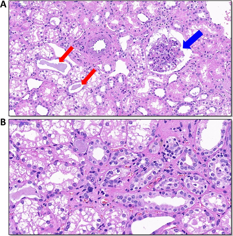
FIG 7.
Histopathology of kidney tissue in monkey M1. Kidney tissue was collected at 25 weeks postinoculation. (A) Protein casts (red arrows) and proliferation of the glomerular mesangium were found (blue arrow). (B) In the renal interstitium, thickening of tubular membranes and inflammatory cell infiltrates were seen.

