FIG 1.
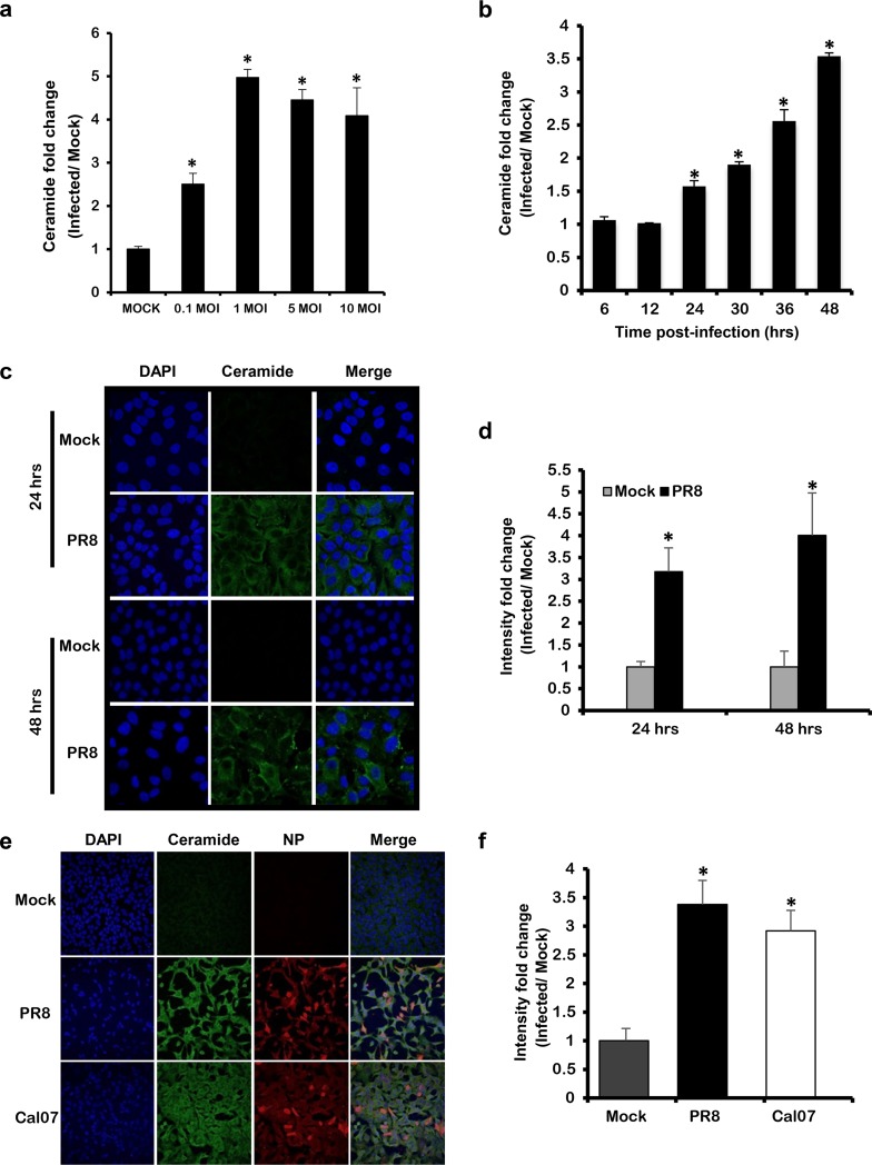
Ceramide accumulates in response to IAV infection in a dose- and time-dependent manner. (a) A549 cells were infected with increasing virus titer of IAV (PR8) for 48 h, or (b, c, and d) with 1 MOI for the indicated time points. (e and f) A549 cells were infected with Cal07 or PR8 at 1 MOI for 48 h. (a and b) Ceramide accumulation was determined using (DGK) assay. For each condition, ceramide accumulation was normalized to total phosphate, and the ceramide fold change was computed with respect to time matching noninfected controls (Mock). (c and e) Ceramide accumulation was detected by confocal microscopy using fluorescent anti-ceramide antibody (green), nuclei were counterstained with DAPI (blue), and IAV was visualized using anti-NP antibody (red) at ×63 (c) and ×40 magnifications. The intensity fold change in ceramide levels between infected cells and time matching Mock was assessed in panels d and f. Statistical significance was determined using the t test (*, P < 0.05) relative to time matching Mock.

