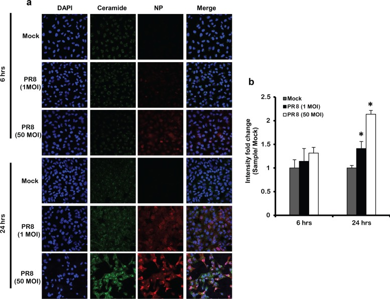
FIG 2.
IAV infection does not induce ceramide accumulation at early viral replication cycles. (a) Ceramide accumulation was detected by confocal microscopy in cells infected by IAV (PR8) using 1 and 50 MOI at the indicated time points (6 and 24 h) or those left noninfected (Mock). Immunostaining was done using anti-ceramide antibody (green), nuclear stain DAPI (blue), and anti-NP antibody (red) at ×40 magnification. (b) The intensity fold change in ceramide levels was assessed between infected cells and time matching Mock, Statistical significance was determined using the t test (*, P < 0.05) relative to time matching Mock.

