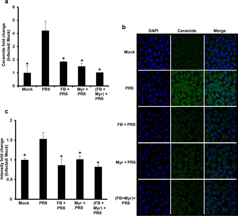
FIG 4.
IAV infection mediates ceramide biosynthesis through the de novo pathway. A549 cells were treated with 50 μM FB and/or 0.1 μM Myr or vehicle for 2 h. The cells were infected with IAV (PR8) at 1 MOI in the presence or absence of the inhibitor(s) or left noninfected. After 48 h, ceramide accumulation was determined by two methods: DGK assay where ceramide fold change was assessed with respect to time matching noninfected controls (Mock) (a) or by confocal microscopy (b and c). Immunostaining was done using anti-ceramide antibody (green) and nuclear stain DAPI (blue) at ×40 for panel b. (c) The intensity fold change between different experimental setups and Mock was measured. Statistical significance was assessed using the t test (*, P < 0.05) relative to IAV-infected, nontreated cells.

