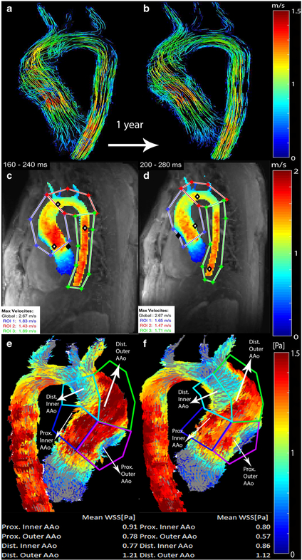
◀Fig. 1.
Comparison of baseline (a, c, e; 16.7 years old) and follow-up (b, d, f; 17.7 years old) scans for a boy with bicuspid aortic valve with right–left commissure fusion pattern. Systolic 3-D streamline flow patterns color-coded by velocity (in m/s) at different time points. a, b Peak systolic velocities were extracted from velocity maximum-intensity projections (c, d) in the ascending aorta, (AAo) arch, and descending aorta. Regional wall shear stress (WSS) values (in pascals, or Pa) were calculated from wall shear stress maps (e, f) for inner proximal, outer proximal, inner distal and outer distal ascending aortic regions

