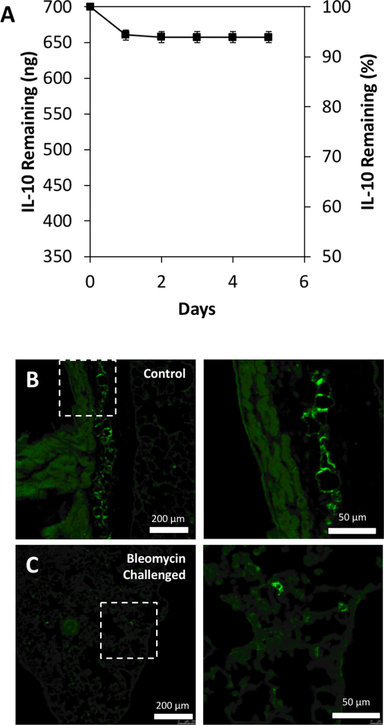
Figure 2. IL-10 immobilization in HH10 and lung deposition of gel.
A) ELISA quantification of IL-10 released from HH-10 formulation over 6 days (n=13). B)-C) Fluorescent microscopy images of lung tissue with fluorescently labeled HH reagent in both healthy (B) and bleomycin-challenged (C) lung tissue, displaying deposition locations of HH reagent via intranasal treatment. Images on right are high magnification of the squared regions.

