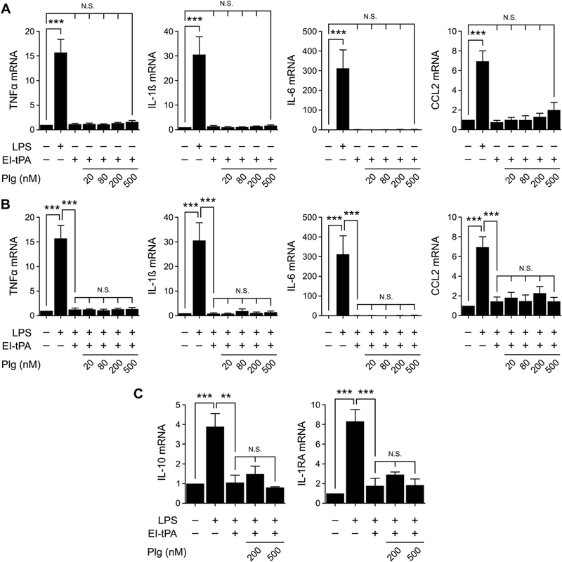
Figure 2. EI-tPA neutralizes the effects of LPS on cytokine expression in BMDMs in the presence of plasminogen.

(A) BMDMs were treated with LPS (0.1 μg/mL) or with EI-tPA (12 nM) alone and in the presence of increasing concentrations (20–500 nM) of plasminogen (Plg) for 3 h. (B) BMDMs were treated with LPS (0.1 μg/ml) alone or with LPS plus EI-tPA (12 nM) in the presence of increasing concentrations of Plg for 3 h. (C) BMDMs were treated with LPS, LPS plus EI-tPA (12 nM), or with LPS plus EI-tPA and Plg (200 or 500 nM). RT-qPCR was performed to compare mRNA levels for TNFα, IL-1β, IL-6, CCL2, IL-10, and IL-1RA (mean ± SEM; n = 5 in panels A and B and 3 in panel C; ***p<0.001, N.S., not statistically significant; one-way ANOVA with Bonferroni’s post hoc test). The presented results show “fold increase” in mRNA expression compared with cultures treated with vehicle (no LPS, EI-tPA, or Plg).
