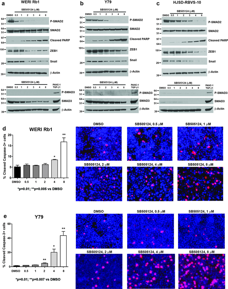
Figure 4. Pharmacological inhibition of the ACVR1C/SMAD2 pathway induces apoptosis and inhibits the expression of EMT markers in retinoblastoma cells.

a-c: Phosphorylation of SMAD2 was reduced in a dose-dependent manner in WERI Rb1 (a), Y79 (b), HSJD-RBVS-10 (c) cells treated with SB505124 for 4 days at the indicated doses, as found by Western blot. Induction of cleaved PARP, indicative of apoptosis, and reduction in Snail and ZEB1 protein levels were also observed, starting at the dose of 2 μM. No phosphorylation of SMAD3 was detected in these lines, while a dose-dependent decrease in the protein levels of SMAD3 was observed in WERI Rb1 and Y79 (Figure 4a-b, bottom panel). PANC-1 cells treated with TGF-β1 at 10 ng/mL for 2 hours were used as positive control for phospho-SMAD3 antibody. d,e: Treatment with SB505124 for 3 days significantly increased apoptosis at 4 and 8 μM in WERI Rb1 (d) and at 2, 4, 8 μM in Y79 (e), compared to DMSO, as determined by immunofluorescence assay using an antibody specific for cleaved caspase-3 (red). P values were calculated using two-sided Student t-test vs DMSO-treated cells. Data are presented as mean + SD. Microphotographs in the right part of the panels are representative images of the immunofluorescence staining. Nuclei were stained with DAPI (blue).
