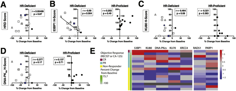
Figure 4. Relationship between H-scores for multiple NHEJ DNA repair pathway proteins and carcinoma shrinkage.
A, relationship between Myriad HRD score and largest percent change in tumor cross sectional area from baseline in target lesions by RECIST [18] in the subset of ovarian cancers for which a numerical HRD score was available (n=13). B-D, IHC staining for 53BP1 (B), KU80 (C), and DNA-PKCS (D) was evaluated to give a modified H-score, which was compared to percent change in tumor cross-sectional area from baseline [18]. HR gene alterations are denoted as mutations (white) or methylation (blue) of BRCA1 (squares), BRCA2 (triangle), or RAD51C (diamond). Symbols that represent two separate patients but are not separable because of overlapping data points are denoted in grey. Ovarian cancers that were stained were sorted into those that were HR deficient (left panels) and those that were not HR deficient (right panels). Because not all ovarian cancers had sufficient sample to assess all antigens, the number of samples analyzed was: 53BP1 (B), HR-deficient n=16, HR-proficient n=16; KU80 and DNA-PKCS (C, D), HR-deficient n=15, HR-proficient n=10. Heat map analysis (E) reflects the H-score for all proteins analyzed, sorted by HR-deficiency and percent change in tumor cross-sectional area from baseline.

