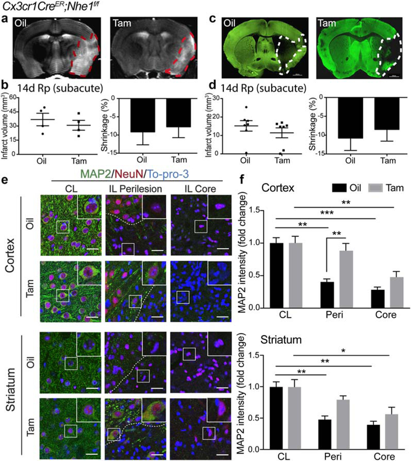
FIGURE 4.

Tam-treated Cx3cr1-CreER;Nhe1f/f mice did not affect subacute infarction but reduced neuronal injury at the subacute phase postischemia. (a) Representative T2-weighted imaging (T2WI) of ex vivo oil- and Tam-treated Cx3cr1-CreER;Nhe1f/f brains at 14 days Rp. (b) Infarct volume and brain atrophy in oil- or Tam-treated Cx3cr1-CreER;NHE1f/f brains at 14 days Rp with T2WI. Data are mean ± SEM. N = 4. (c) Representative MAP2 staining of oil- and Tam-treated Cx3cr1-CreER;Nhe1f/f brains at 14 days Rp. (d) Infarct volume and brain atrophy in oil- or Tam-treated Cx3cr1-CreER; NHE1f/f brains at 14 days Rp with MAP2 staining. Data are mean ± SEM. N = 7. (e) Representative immunofluorescent images of MAP2, NeuN, and To-pro-3 staining in the CL, IL perilesion and IL core areas in the cortex and striatum of the oil- and Tam-treated Cx3cr1-CreER;Nhe1f/f mice at 14 days post-tMCAO. (f) Quantitative analysis of MAP2 intensity in the CL, IL perilesion and IL core areas in the cortex and striatum of the oil- and Tam-treated Cx3cr1-CreER;Nhe1f/f mice at 14 days post-tMCAO. N = 3. *p < .05, **p < .01, ***p < .001 [Color figure can be viewed at wileyonlinelibrary.com]
