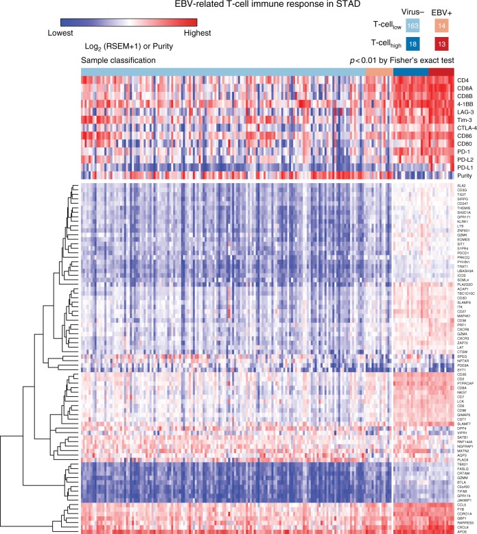
Fig. 4.
The supervised clustering of T-cell genes in EBV-positive and virus-negative STAD tumors. In the upper panel, we show the expression of T-cell markers such CD4, CD8 and immune checkpoint genes PD-L1, PD-L2, PD-1, CD80, CD86, CTLA-4, Tim-3, LAG-3, and 4-1BB. Tumors are clustered to four different groups based on the T-cell and virus statuses, i.e., Virus-/T-celllow, EBV+/T-celllow, Virus-/T-cellhigh, and EBV+/T-cellhigh from left to right

