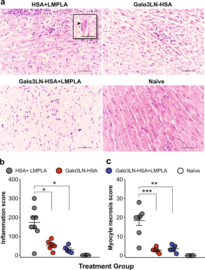
Fig. 6.
Histopathology analysis. a Micrographs of heart sections harvested at the endpoint and stained with hematoxylin and eosin. Inset, arrowhead indicates an amastigote nest. b Myocardial inflammatory and mononuclear cell infiltrates were manually scored. c Myocyte necrosis was manually counted in 25 fields (×400). Magnification ×40, bar 100 μm. Statistical significance was calculated by unpaired two-tailed Mann–Whitney test, comparing Galα3LN-HSA+/−LMPLA group to HSA+LMPLA group. p values indicate the significance *p < 0.05; **p < 0.01; ***p < 0.001

