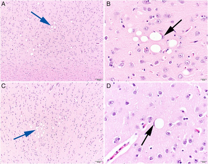
Figure 1.

Histopathological changes in the cerebral cortex of a 3‐year‐old (A and B) and a 10‐year‐old (C and D) Boerboel with focal epilepsy. Individual and clusters of neurons reveal single, variably sized optically empty vacuoles in the perikaryon (blue arrows). Regularly compression and displacement of the nucleus are visible (black arrows). H&E stain, size bar 100 μm (A and C) and 20 μm (B and D)
