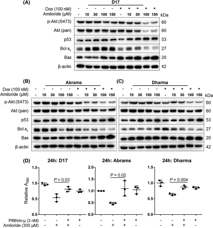
Figure 4.

Western blot of proteins involved in p53 and Akt signaling in (A) D17, (B) Abrams, and (C) Dharma cells. (D) Viability after pharmacological inhibition of p53 translocation to the mitochondria. Cells were treated with pifithrin‐μ (3 nM) alone or in combination with 300 μM amiloride for 24 hours. Cell viability was measured with the crystal violet assay. Means were presented with SD, and significance reported at *P < .05
