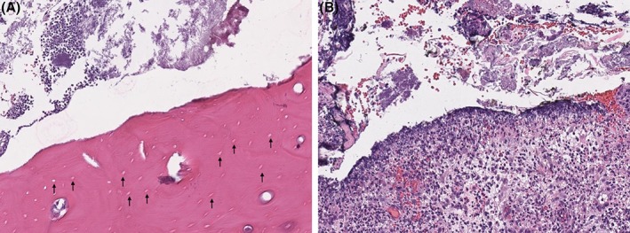
Figure 3.

Histopathology of exposed maxillary bone and adjacent soft tissue. A, Mandibular bone with black arrows noting absence of osteocytes within numerous lacunae, consistent with osteonecrosis. B, Marked suppurative to histiocytic ulcerative gingivitis with mixed bacteria
