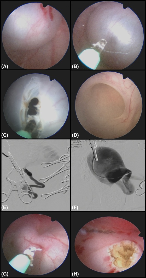
Figure 2.

Endoscopic and fluoroscopic images of a 2‐month‐old male intact Labrador Retriever with severe left hydroureter and hydronephrosis and an imperforate orthotopic ureterocele on the left and an ectopic ureter on the right. This procedure was performed from an antegrade cystoscopy approach owing to the small size of the canine pelvic urethra limiting the ability to perform percutaneous perineal access cystourethroscopy. The dog is in dorsal recumbency. A, A large fluid‐filled structure covering the left ureteral orifice. There is no orifice seen. B, A diode laser is used to puncture the fluid‐filled structure. C, The orifice in the structure is expanded with the laser and then the scope is used to explore the structure (D). D, The ureteral opening is seen inside this fluid‐filled structure and is largely dilated. E, A digital subtraction contrast study is done using as a retrograde ureterogram up the ureteral orifice showing the severe hydroureter and hydronephrosis. F, Contrast is injected into the cyst after it is opened using a ureteral catheter, and the cyst and wall can be seen inside the urinary bladder as contrast refluxes retrograde up the ureteral orifice. G, Laser ablation of the contralateral ectopic ureter from an antegrade approach as the ureteral orifice could not be cannulated retrograde owing to the small size of the canine penis at this young age. H, Endoscopic image after both orifices are open and the filled cystic structure was ablated
