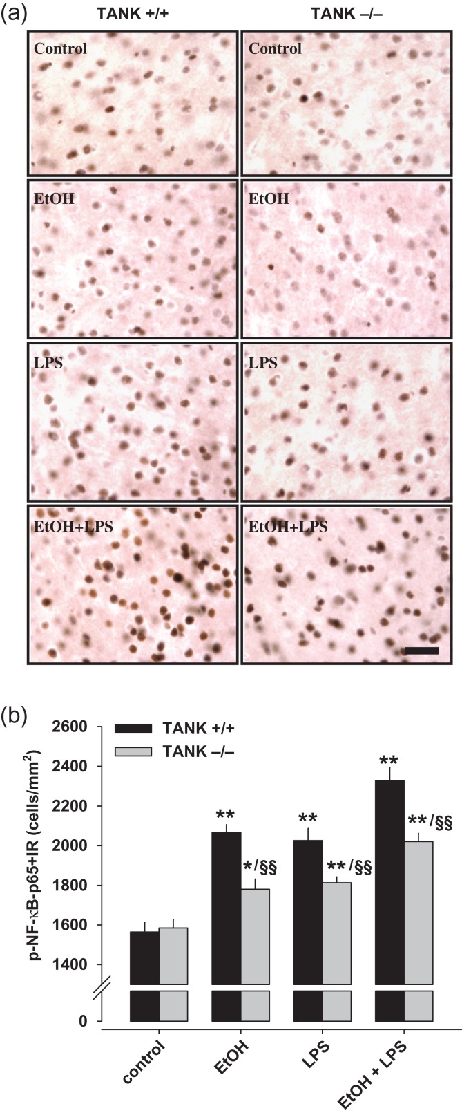
Figure 6.
TANK controls neuroimmune activation after alcohol and/or lipopolysaccharide (LPS) in the insular cortex of mice. (a) Images of p-NFκB-p65+immunoreactive cells (+IR) of the anterior insular cortex of tank KO and WT mice after alcohol (5 g/kg, i.g., 10 days) and LPS (3 mg/kg, i.p.) treatment (scale bar = 30 μm). (b) Quantification of p-NFκB-p65+IR cells. BioQuant image analysis shows that cell number of p-NFκB-p65+IR was significantly increased after 10 days of alcohol (i.g.) or LPS alone. Pretreatment of alcohol significantly enhances LPS-induced p-NFκB-p65 immunoreactivity in both WT and tank KO mice (ANOVA: P < 0.01). Anterior insular cortex of tank KO mice shows significantly decreased p-NFκB-p65+IR cells after alcohol and/or LPS compared with WT mice (t-test; *P < 0.05, **P < 0.01 vs. control treatment; §§P < 0.01 vs. WT mice with same treatment).

