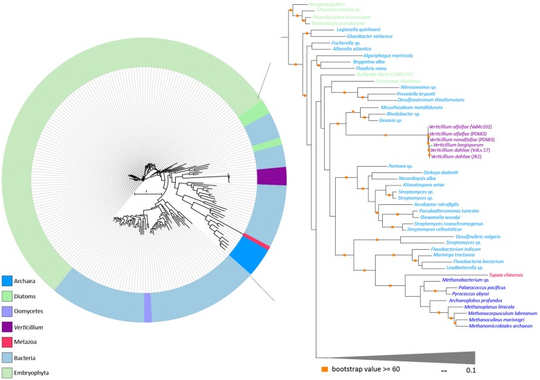
Fig. 4.
—Evolutionary relationship of HGT-2 homologs. Protein sequences of HGT-2 homologs were aligned (using MAFFT) and the resulting alignment was used to infer a maximum-likelihood phylogeny (using RAxML). Different colors depict different groups or species. The phylogeny suggests that HGT-2 is transferred from a bacterial species. A more detailed part of the tree that contains Verticillium species is shown on the right. Orange squares indicate branches with bootstrap values ≥60.

